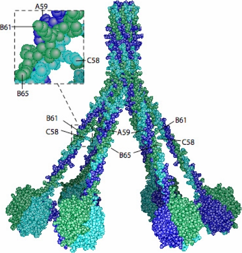
Fig. 10.
Three-dimensional model of human C1q highlighting positions of unmodified lysine residues. The model (2) was assembled as described previously (50). The C1q chains are colored dark blue (A), green (B), and light blue (C). The positions of the side chains of Lys59 A, Lys61 B, and Lys58 C (unmodified) and of Lys65 B (hydroxylated) are shown.

