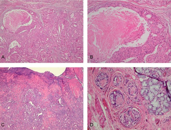
Figure 3.
Microscopic findings. (A) Pleomorphic adenoma in a chondromyxoid, fibrotic and sclerotic stroma with the malignant areas of carcinoma component consisting epithelial clusters with necrosis. (H&E, 40×), (B) Carcinoma component is composed of cells with pleomorphic nucleus, mitotic activity and comedo-like necrosis. (H&E, 100×), (C) The carcinoma ex pleomorphic adenoma consisting benign pleomorphic adenoma and poorly differentiated carcinoma with the ulcerative epithelial surface. (H&E, 40×), (D) Tumor cells presented the perineural invasion (H&E, 400×).

