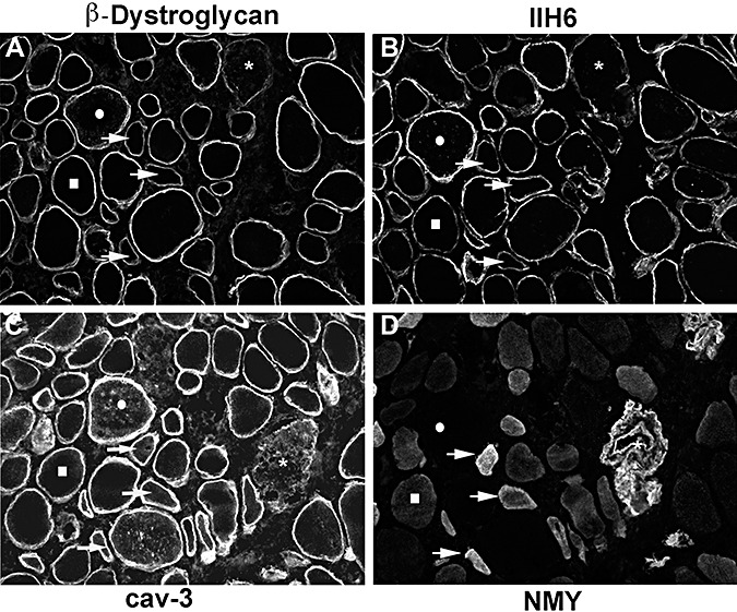
Figure 6.

The biopsy of P11 showed the mildest reduction in IIH6 labeling. The labeling of individual fibers with antibodies to β‐dystroglycan (A), IIH6 (B), caveolin‐3 (cav‐3; C) and neonatal myosin (NMY; D) is compared. A few fibers with reduced IIH6 labeling and normal β‐dystroglycan immunoreactivity were seen (squares). Some fibers showed reduced IIH6 and β‐dystroglycan and were positive for the neonatal isoform of myosin so they may represent regenerating fibers (arrows). Fibers with low β‐dystroglycan, IIH6 and reduced sarcolemmal cav‐3 labeling may reflect sarcolemmal damage (asterisks). A population of fibers has marked IIH6 and β‐dystroglycan labeling on the cell surface and intracellular labeling of cav‐3 but no neonatal myosin labeling (circles).
