
Figure 3.
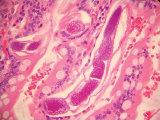
High power microscopic view showing larvae embedded within the small bowel villi. Notice the dark purplish organs surrounded by translucent sheath (coat) (hematoxylin and eosin ×400).
Official websites use .gov
A
.gov website belongs to an official
government organization in the United States.
Secure .gov websites use HTTPS
A lock (
) or https:// means you've safely
connected to the .gov website. Share sensitive
information only on official, secure websites.
High power microscopic view showing larvae embedded within the small bowel villi. Notice the dark purplish organs surrounded by translucent sheath (coat) (hematoxylin and eosin ×400).