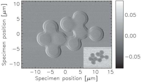
FIG. 2.

Horizontal component of the DPC signal Sx obtained from a cluster of 5-μm-diameter polystyrene spheres. The step within the spheres is due to the presence of residual solution. Inset: absorption contrast image obtained from the sum of all detector segments. The peak specimen absorption is about 7%. The scan was recorded in 401 by 301 steps of 75 nm using a 5-ms dwell.
