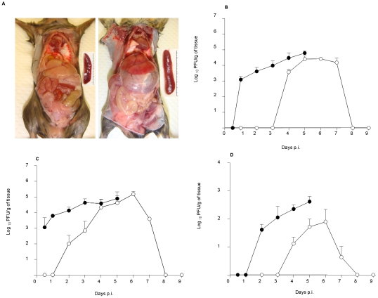
Figure 4. Pathology and virus titres in the liver, spleen and brain of D2Y98P-infected mice.
(A) Mice were ip. infected with 107 PFU of D2Y98P, and were sacrificed at moribund state and perfused extensively with PBS. Representative gross appearance of organs in the intraperitoneal cavity of uninfected (left panel) and ip. infected (right panel) mice. Insets highlight the difference in the spleen size between both animal groups. Virus titres were determined in the liver (B), spleen (C) and brain (D) from AG129 mice ip. infected with 107 (black circle) or 104 (open circle) PFU of D2Y98P virus. Results are expressed as log10 [mean ± SD] in PFU per gram of tissue. Five mice per time point per group were assessed. Results are representative of 2 independent experiments.

