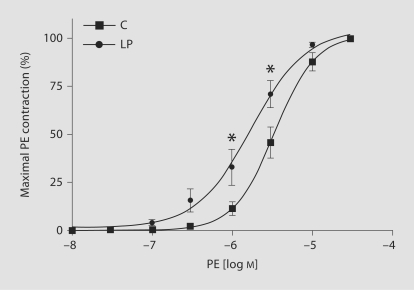
Fig. 3.
PE-induced contraction in mesenteric arterial rings of C and LP rats. Endothelium-intact mesenteric arterial rings were incubated in normal KPSS and then stimulated with increasing concentrations of PE. PE contraction was measured and presented as percentage of maximum PE contraction (semilog plot). Data points represent means ± SEM of measurements in 10–12 mesenteric arterial rings from 5–6 rats of each group. * p < 0.05 compared with C rats.

