
FIGURE 6. Space-filling representation of the environment around the guanosine nucleophile.
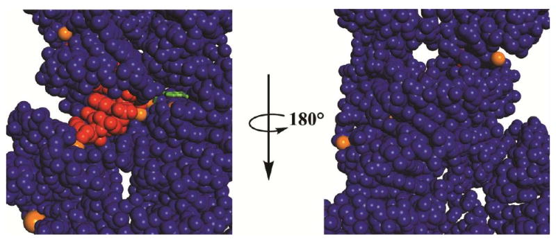
The guanosine nucleophile is in green, the oligonucleotide substrate in red, metal ions in orange, and the ribozyme in blue. The guanosine nucleophile is shown as a stick representation and all other atoms as space filling. Only the central portion of the Azoarcus structural model is shown (3bo3, ref. 41).