
Figure 4.
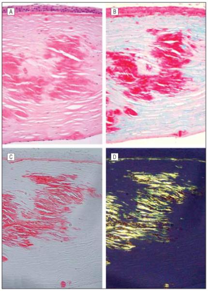
Histopathologic examination of the excised corneal button from the right eye of the proband. Large eosinophilic deposits are noted in the anterior, mid, and posterior corneal stroma (A, hematoxylin-eosin, original magnification ×200). The deposits noted stain brightly with Masson trichrome (B, original magnification ×200) and Congo red (C, original magnification ×250). The Congo red-stained section demonstrates dichroism with the use of polarizing filters (D), produced by the presence of amyloid fibrils in the stromal deposits.