
Figure 5.
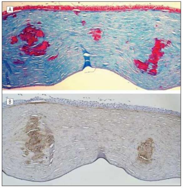
Histopathologic and immunohistochemical analysis of the excised corneal button from the left eye of the proband. A, Large stromal deposits stain bright red with Masson trichrome (original magnification ×200). B, The same deposits also stain with an antibody to transforming growth factor β–induced (TGFBI) protein, indicating that the deposits represent focal aggregations of the protein product of the TGFBI gene (original magnification ×200).