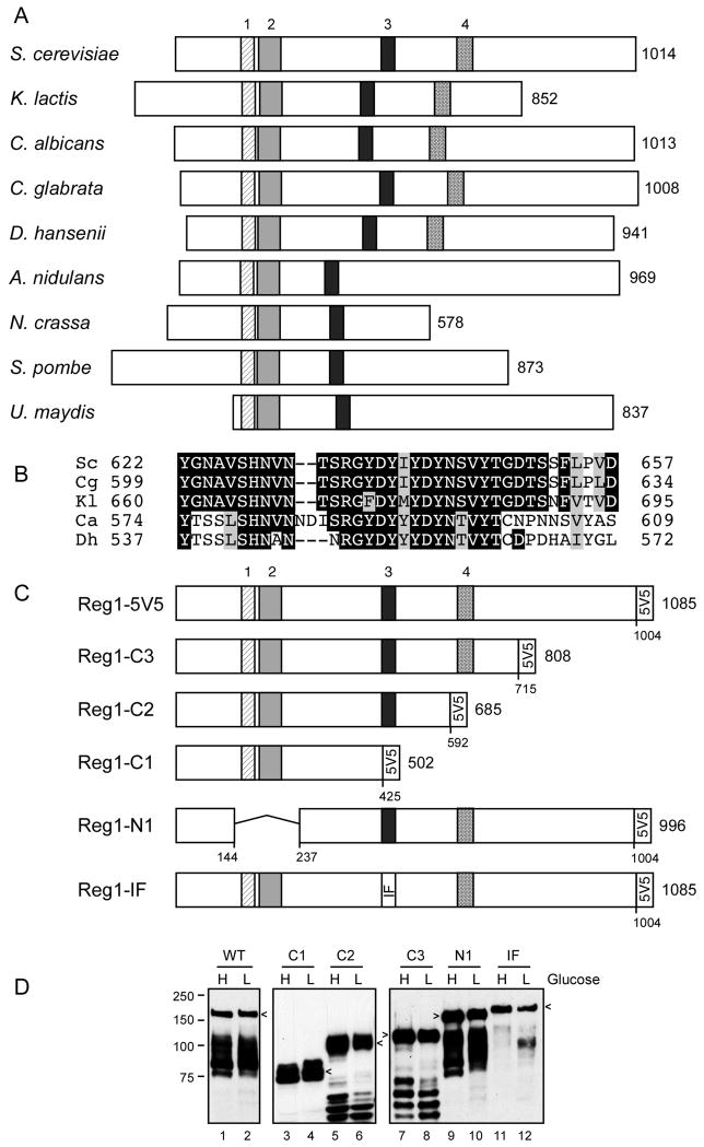
Fig. 1.
Conserved domains of the Reg1 protein. (A) Schematic representation of Reg1 orthologs from nine fungal species. The number of amino acids in each protein is shown on the right. The size and position of four conserved blocks of sequences are drawn to scale. Blocks 1 through 3 were previously noted [17]. (B) Multiple sequence alignment of conserved block 4. (C) Reg1 deletion constructs and the double point mutation (IF: I466M, F468A) are shown with the 5 tandem copies of the V5 epitope on the C-termini. The total length of each protein and deletion junctions are shown. (D) Western blot of Reg1-V5 proteins isolated from cells grown in high (H) or low (L) glucose. Arrows indicate mobility of the full-length protein. Faster migrating species represent proteolytic fragments.

