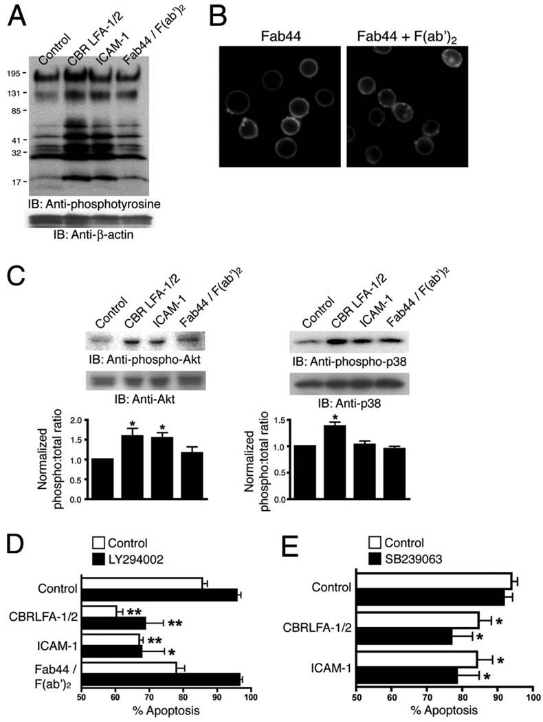
FIGURE 1.
A and C, PMN were treated at 37°C for 15 min with 10 µg/ml CBR LFA-1/2 or ICAM-1, both in the presence of 1 mM MnCl2 or with anti-αM Fab (Fab44) and secondary F(ab′)2 to induce Mac-1 clustering. For cells treated with CBR LFA-1/2, anti-αM Fab fragments at a concentration of 50 µg/ml were used to block potential ligand binding. In A, immunoblots of whole-cell lysates were probed for the presence of tyrosine-phosphorylated proteins and for β-actin as a protein loading control. In C, immunoblots were probed for phosphorylated (phospho-p38; pThr180/pTyr182) and total p38, or for phosphorylated (phospho-Akt; pThr308) and total Akt. Western blot results from two experiments were quantified using ImageJ software and were normalized according to the control treatment. B, Unstimulated PMNs were incubated at 37°C for 15 min with FITC-conjugated anti-αM Fab44 (5 µg/ml) in the absence (left) or presence (right) of secondary F(ab′)2 (2.5 µg/ml) to induce Mac-1 clustering. D and E, PMN were treated with 1 mM MnCl2, 10 µg/ml CBR LFA-1/2 Fab and 10 µg/ml anti-αM Fab (Fab44), 1 mM MnCl2 and 200 µg/ml ICAM-1, or 5 µg/ml anti-αM Fab (Fab44) and 2.5 µg/ml secondary F(ab′)2 to induce Mac-1 clustering. Cells were pretreated for 15 min with or without 40 µM LY294002 or 2 µM SB239063. After incubation at 37°C for 16 h, cells were labeled with FITC-conjugated annexin V to detect apoptotic PMNs. Data are means ± SEM from three (D) or five (E) experiments performed in duplicate, and are expressed as percent of annexin V-positive cells. Significantly different from control (**, p < 0.01; or *, p < 0.05).

