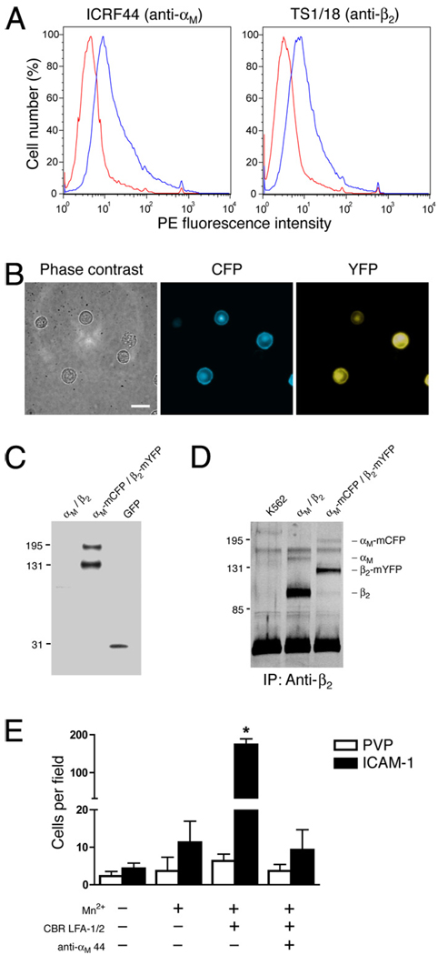
FIGURE 6.
A, K562 cells expressing αM-mCFP and β2-mYFP were labeled with anti-αM ICRF44 mAb, anti-β2 TS1/18 mAb, or IgG1 isotype mAb (red trace), followed by PE-conjugated secondary Abs. Cells were analyzed by flow cytometry. B, K562 cells expressing αM-mCFP and β2-mYFP were visualized under phase contrast and immunofluorescence microscopy. C, Whole-cell lysates from K562 cells expressing αM-mCFP and β2-mYFP were analyzed by immunoblotting using an anti-GFP polyclonal Ab. D, Mac-1 was immunoprecipitated from K562 cells, K562 cells expressing wild-type αM and β2, and K562 cells expressing αM-mCFP and β2-mYFP using the anti-β2 CBR LFA-1/2 mAb. Proteins were separated on a 7.5% SDS-PAGE gel and then analyzed by silver staining. E, K562 cells expressing αM-mCFP and β2-mYFP were allowed to adhere to tissue culture plastic coated with ICAM-1 or PVP, as a control, in the presence or absence of 1 mM MnCl2, 10 µg/ml CBR LFA-1/2 mAb, and anti-αM clone 44 mAb. After a 30-min incubation, nonadherent cells were washed away, and the number of adherent cells per field was counted. Data are presented as mean ± SEM from one of three independent experiments. *, Significantly different from control (p < 0.01).

