Figure. 1.
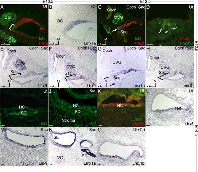
Expression of LIM-HDs in the developing inner ear: E10.5 (A,B), E12.5 (C-H) and E14.5 (I-O). Each stage was labeled with sidebars. A: ISL1 labeling in the region of future sensory epithelia (bracket) was distinct from primarily non-sensory PAX2 expression (red) in the medial portion of otocyst (Ot). OG, otic ganglions. B: In adjacent section, prominent Lmx1a expression was in a region overlapping with PAX2 expression. C: At E12.5, ISL1 expression was prominent in primordial cochlea (coch), cochleovestibular ganglions (CVG) and the developing saccular sensory epithelia, which overlapped slightly with PAX2 expression (arrow). D: Labeling with antibodies against LHX3 and POU4F3 showed immunoreactivities in nascent utricular hair cells. Most hair cells were LHX3 and POU4F3 double-positive (arrows to show example), whereas occasional early hair cells were only positive for POU4F3 (arrowhead). E-H: Expression of Lhx5, Lhx9, Lmx1a and Lmx1b in the inner ear at E12.5. Among them, only Lhx5 was expressed in the primordial cochlea (Fig.1E). Lmx1a was detected in the non-sensory epithelial region, but undetectable in the sensory epithelium positive for ISL1 (arrows, comparing to 1C) or in the cochleovestibular ganglions (Fig.1G). The sensory epithelial region of saccule is indicated by brackets, which correspond with ISL1 staining in adjacent section (data not shown). I: ISL1 expression in utricle at E14.5 was mainly in supporting cells (SC), with little expression in hair cells (HC). J: ISL2 expression was higher in saccular hair cells, weak in supporting cells and stroma. K: LHX3 expression was only in the utricular hair cells, coinciding with Myosin VIIa (MYO7A). L, M and O: Lhx5, Lhx9 and Lmx1b were detected in the sensory epithelial cell region. N: Robust Lmx1a expression was restricted to non-sensory epithelia including endolymphatic duct (ED), and was excluded in the vestibular ganglions (VG). Ut, utricle; Sac, saccule; Cri, crista; SE, sensory epithelia. Scale bar=20μm.

