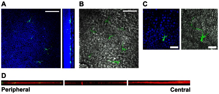
Figure 2. CD11c-expressing dendritic cells reside in the basal epithelium.
A–C) Whole corneas from Balb/c mice expressing EGFP from the CD11c promoter (green) were stained with DAPI (blue) and imaged by confocal microscopy. A) Left: Representative slice through basal epithelial layer of the paracentral region of the cornea. Right: Images were reconstructed in the yz plane to show the position of EGFP+ dendritic cells in the basal layer of the corneal epithelium demarked by the heavy density of cell nuclei. Bar, 75 μm. B) Same optical section as in A (left) with green DCs overlaid on corresponding differential interference contrast (DIC) image. Bar, 75 μm. C) High magnification image of a DC in the basal epithelial layer. Bar, 25 μm. D) Corneal epithelial sheets were dissociated from whole corneas by treatment with EDTA, stained for laminin, a marker of the epithelial basement membrane, and imaged by confocal microscopy. The images were reconstructed in the xz plane and form a montage from the peripheral cornea on the left to the center of the cornea on the right, with the ocular surface above the images.

