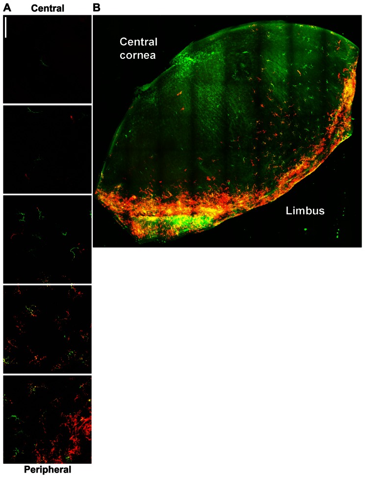
Figure 3. MHC class II expression on corneal DCs.
Corneal whole mounts from mice expressing EGFP from the CD11c promoter were stained for MHC class II and imaged by confocal microscopy. A) Montage of images showing CD11c promoter activity (green) and surface MHC class II expression (red) from central (top) to peripheral (bottom) cornea. Bar, 75 μm. B) Montage of images from a large portion of cornea with CD11c promoter in green and MCH call II expression in red.

