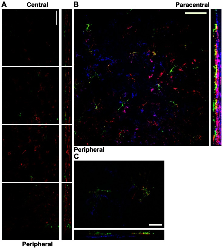
Figure 4. Stratification of APCs within the normal cornea.
Corneal whole mounts from mice expressing EGFP from the CD11c promoter were stained for CD11b alone (A) or CD11b and MHC class II (B and C) and imaged by confocal microscopy. A) Corneal whole mounts from mice expressing EGFP from the CD11c promoter (green) were stained for surface CD11b expression (red). Montage of images (xy plane on the left; yz plane on the right with epithelium toward the left) from the corneal center (top) to periphery (bottom). Bar, 75 μm. B and C) Corneal whole mounts from mice expressing EGFP from the CD11c promoter (green) were stained for surface MHC class II (red) and CD11b (blue). B) Representative low-magnification image of the peripheral (lower left) and paracentral (upper right) cornea. Bar, 100 μm C) Representative high-magnification image from the peripheral/paracentral region. Bar, 25 μm.

