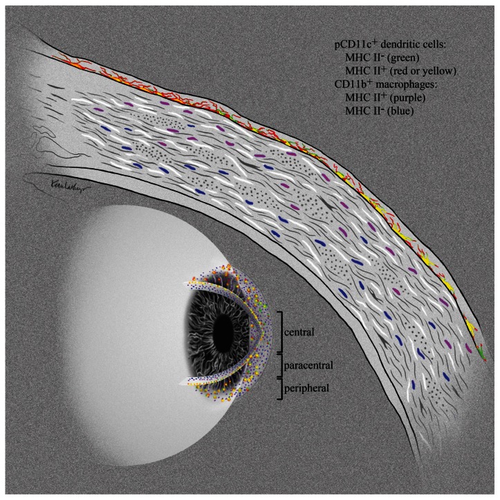
Figure 5. Stratification of APCs within the normal cornea.
Schematic diagram showing the stratification of APCs within the normal cornea. EGFP+ (green) DCs that co-express MHC class II (red or yellow) reside within the epithelial basement membrane extending dendrites toward the ocular surface. Below the epithelial basement membrane, MHC class IIdim CD11b+ putative macrophages (purple) sit in the anterior stroma, while MHC class II− CD11b+ cells (blue) fill the remainder of the posterior stroma. This unique stratification of APCs suggests progression from an antigen presentation function at the exposed corneal surface to an innate immune barrier function deeper in the stroma.

