Figure 2.

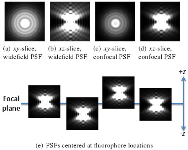
PSFs from widefield fluorescence microscope (a-b) and confocal fluorescence microscope (c-d) calculated with XCOSM [MC02]. Intensities are scaled to emphasize the overall shape of the PSFs. (e) Model of fluorescence microscope image formation. PSFs centered on fluorophores intersect the focal plane. Summing the PSF xy-slices at the intersections yields the simulated fluorescence image.
