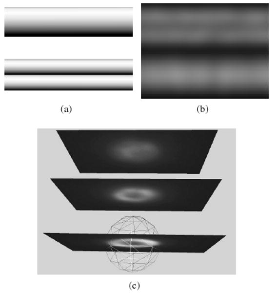
Figure 3.

Examples of unintuitive images in fluorescence microscopy. (a) Models of surface-labeled small tubes. The side-by-side tubes are half the diameter of the larger tube. (b) Simulated noise-free fluorescence image of the tubes convolved with a calculated widefield PSF generated by XCOSM [MC02]. The two tubes can easily be misconstrued as a single tube while the single tube appears as two tubes. (c) A surface-labeled bead (in wireframe) with 1 micron radius superimposed with experimental images at focal planes with 2 micron spacing. The top-most image could be interpretted as the top of the sphere, but it is four times further away from the sphere center than the true top.
