
Fig. 1.
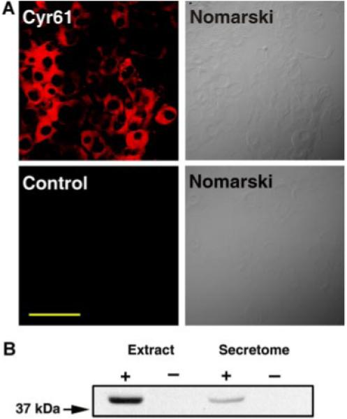
The expression of Cyr61 in MSCs. A: mMSCs were immunostained with (upper left part) or without (lower left part) anti-Cyr61 antibody, as described in Experimental Procedures Section. The corresponding Nomarski micrographs are shown in the right parts. Scale bar, 100 μm. B: Western blot analysis of mMSC cellular extracts and secretome with Cyr61 antibody. Left lane, cellular extracts (66 μg); second lane, extraction buffer (negative control); third lane, secretome (66 μg); right lane, plain-DMEM (negative control). Note that Cyr61 polypeptides are detected in both cell extracts and secretome. [Color figure can be viewed in the online issue, which is available at www.interscience.wiley.com.]