
Fig. 2.
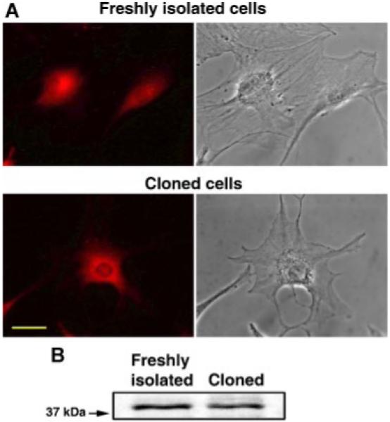
The expression of Cyr61 in freshly isolated and cloned MSCs. A: Freshly isolated (upper left part) and cloned (lower left part) MSCs were immunostained with anti-Cyr61 antibody, as described in Experimental Procedures Section. The corresponding phase contrast micrographs are shown in the upper and lower right parts, respectively. Scale bar 40 μm. B: Western blot analysis of freshly isolated and cloned MSCs with Cyr61 antibody. Note that Cyr61 polypeptides are detected in both freshly isolated and cloned MSC cells. [Color figure can be viewed in the online issue, which is available at www.interscience.wiley.com.]