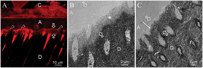
Fig. 3.
Remineralization was most frequently observed along the base of the hybrid layers. C: composite; A: adhesive; Between open arrows: hybrid layer; D: intertubular dentin, T: dentinal tubule. A. CLSM image of a specimen that had been immersed in the biomimetic remineralization medium for 6 months. Fluorescence was predominantly identified from the surface of the hybrid layer (open arrowheads) and from the dentinal tubules beneath the hybrid layer. Quenching of the fluorescence could be seen along the basal portion of the hybrid layer. B. TEM image taken from a specimen slab that had been immersed in the biomimetic remineralization medium for 2 months, showing an initial stage of remineralization that originated from the base of the hybrid layer. Remineralization was not very intense at this stage, as indicated by the less electron-dense nature of the remineralized part of the hybrid layer (asterisk). C. TEM image taken from the 6-month old specimen in Fig. 3A depicting a more advanced stage of remineralization. Remineralization stopped at approximately 1–2 μm from the dentin surface (arrow). The remineralized part of the hybrid layer exhibited a similar mineral density as the underlying unaltered mineralized intertubular dentin.

