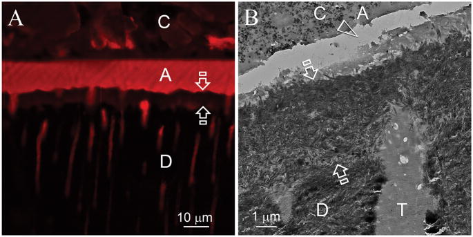
Fig. 5.
In some 4–6 month specimens, extensive remineralization that spanned across the entire thickness of the hybrid layers could be identified. C: composite; A: adhesive; Between open arrows: hybrid layer; D: intertubular dentin, T: dentinal tubule. A. CLSM image of a specimen that had been immersed in the biomimetic remineralization medium for 4 months. Quenching of the fluorescence derived from Rhodamine B was evident throughout the entire hybrid layer (between open arrows). B. TEM image of the same specimen. Heavy remineralization could be seen throughout the hybrid layer. The artifactual gap (open arrowhead) within the adhesive layer was created during ultramicrotomy.

