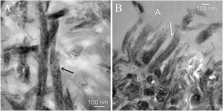
Fig. 6.
High magnification TEM micrographs showing intrafibrillar remineralization of collagen fibrils within the hybrid layer. A. This view was taken from the base of the hybrid layer from a specimen that exhibited an early stage of remineralization. Those collagen fibrils that were better infiltrated by the adhesive did not remineralize and appeared electron-lucent. An ordered arrangement of apatite platelets could be identified within those collagen fibrils that remineralized (arrow). The rationale for the use of biomimetic analogs is to generate apatite nanocrystals that are small enough to fit into the gap zones of the collagen molecules (i.e. intrafibrillar remineralization). B. This view was taken from the dentin surface of a more heavily remineralized specimen. Interfibrillar spaces did not contain minerals as they were filled with adhesive resin. Cross-sections of the remineralized collagen fibrils (open arrowheads) clearly showed that they were fully packed with intrafibrillar minerals. Collagen fibrils along the dentin surface had a shag carpet-like appearance and were partially unraveled along their severed ends. Even these unraveled collagen fibrils were heavily remineralized with apatite nanocrystals (arrow).

