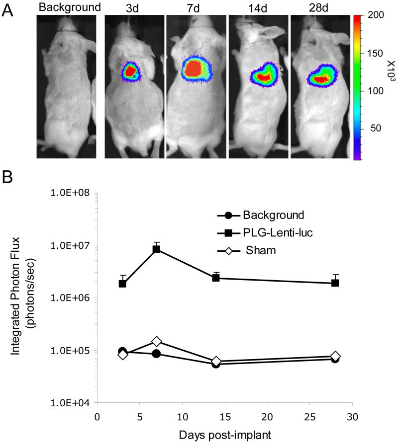
Fig. 6. In vivo tranduction by lentivirus-lyophilized PLG scaffold.
(A) Bioluminescence imaging and quantification of firefly luciferase expression for 4 weeks following subcutaneous implantation of lentivirus immobilized PLG scaffolds. Lentivirus expressing luciferase (Lenti-luc, 3 × 10⁸ LP) was lyophilized onto the unmodified PLG scaffold. (B) Integrated light flux (photons/sec) as measured using constant-size regions of interest over the implant site (n = 4 for day 3, n = 7 for day 7, n = 3 for day 14 and n = 3 for day 28 for experimental and background data). Scaffolds lyophilized with lentivirus expressing luciferase (■), background, (●), and sham operation (◊, n = 1). Values are mean ± S.E.M.

