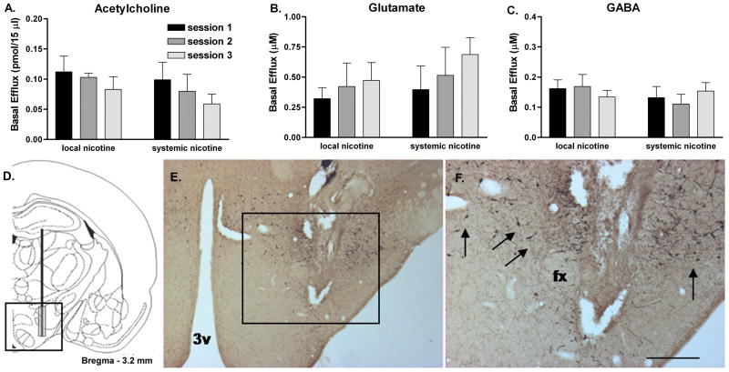
Figure 1.
Mean raw basal LH/PFA efflux values (uncorrected for probe recovery) for ACh (A), glutamate (B) and GABA (C) during the three systemic or local nicotine treatment sessions. Basal efflux was not significantly altered for any of the three neurotransmitters as a function of microdialysis session or route of nicotine treatment. D. Coronal hemisection schematic of probe placement (modified from Paxinos and Watson, 1997) in LH/PFA. E. Representative low-magnification photomicrograph of probe tract in the hypothalamus. F. Higher-magnification of the area outlined in (E) demonstrating the microdialysis probe tract surrounded by orexin-immunoreactive neurons (arrows). Abbreviations: 3v, third ventricle; fx, fornix; scale bar in (F) represents approximately 500μm. Error bars represent SEM.

