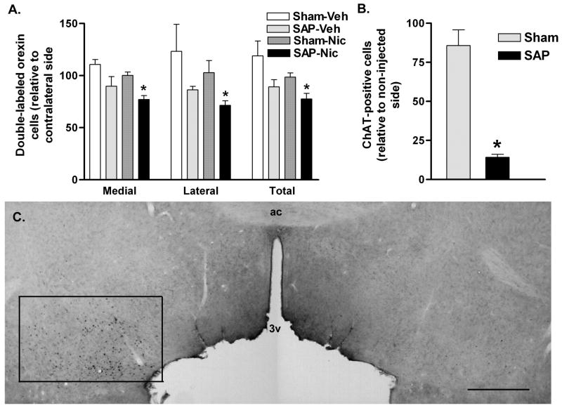
Figure 6.
Activation of orexin neurons by nicotine following basal forebrain cholinergic lesions. A. Double-labeled (Fos/orexin) neurons on the injected side relative to the contralateral hemisphere in control (Sham) or 192 IgG-saporin (SAP) basal forebrain-lesioned animals treated acutely with systemic vehicle (Veh) or nicotine (Nic). 192 IgG-saporin lesions resulted in a significant reduction in nicotine-activated orexin neurons ipsilateral to the lesion site. This attenuation was seen in both medial and lateral sectors of the LH/PFA. *P < 0.05 vs. Sham-Nic. B. 192 IgG-saporin (SAP) treatment resulted in a greater than 80% loss of ChAT-immunoreactive cells in the basal forebrain relative to the non-lesioned hemisphere. No significant loss of cholinergic cells was seen in the sham-lesioned animals. *P < 0.05 vs. Sham. C. Immunohistochemistry for ChAT in the basal forebrain of a unilaterally-lesioned animal. The non-infused (left) hemisphere is characterized by the presence of numerous ChAT-immunoreactive cells in basal forebrain regions encompassing the substantia innominata, ventral pallidum and horizontal limb of the diagonal band of Broca, as indicated by the rectangle. The 192 IgG-saporin-infused (right) hemisphere is largely devoid of ChAT-immunoreactive neurons. Abbreviations: ac, anterior commissure; 3v, third ventricle. Scale bar equals approximately 1 mm.

