
Fig. 3. A hypersomnia case with hypothalamic tumor and low hypocretin level (167 pg/ml).
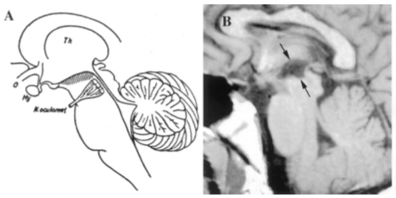
A 23 year-old man who developed narcolepsy-cataplexy after a large hypothalamic stroke. His lesion included much of the hypothalamic region in which orexin is produced. (A) Diagram of a sagittal section of the brain adapted from von Economo 1; the hatching highlights those regions in which inflammatory lesions produced hypersomnia. (B) T1-weighted sagittal and horizontal MR images demonstrating ex vacuo changes (between arrows) in the posterior hypothalamus and rostral midbrain of the patient with secondary narcolepsy. Th = thalamus; o = optic nerve; Hy = hypothalamus; N. oculomot = occulomotor nerve. A permission from ((Scammell et al., 2001)).