
Figure 11.
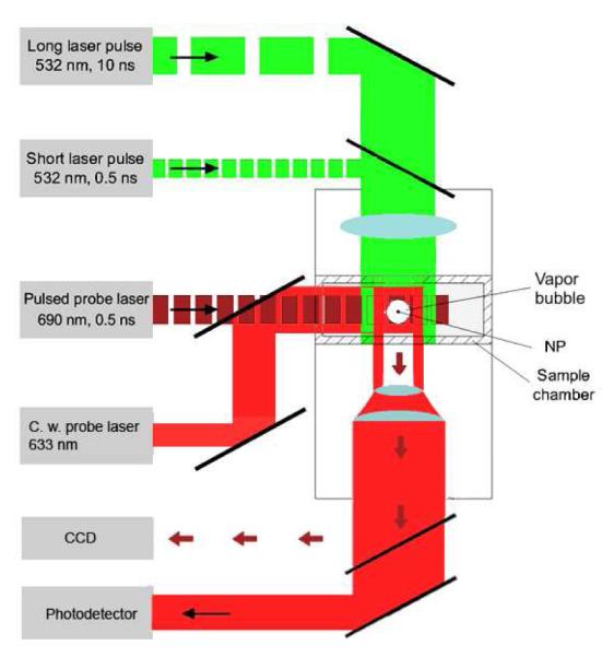
Experimental setup: single gold NPs in water were placed in the sample chamber mounted on the microscope stage; bubble generation was provided by focused single pulses (532 nm, 05 ns or 10 ns); a pulsed probing laser (690 nm, 05 ns) provided time-resolved optical scattering imaging of bubble and a continuous probing laser (633 nm, 1 mW) provided the monitoring of the integral optical scattering of bubble.