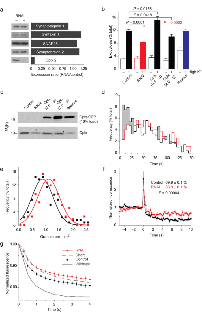
Figure 6.
NPY-mRFP exocytosis in cells depleted of cplx. (a) RNAi-mediated silencing of cplx expression. Cells cotransfected with GFP and a plasmid encoding short hairpin RNA (shRNA) targeting cplx 2 were isolated by fluorescence activated cell sorting and analyzed by immunoblotting. (b) Fluorimetric assay of NPY-mRFP released by cells after a 45-minute incubation at low or high [K+] (n = 15). In addition to 0.5 µg of NPY-mRFP plasmid (control), cells were transfected with 1 µg shRNA (RNAi), 0.5 or 2.0 µg cplx-GFP, or shRNA and 0.5 µg cplx-GFP plasmids (rescue). (c) Immunoblot analysis of the transfection conditions in b. (d–g) TIRFM imaging of NPY-mRFP exocytosis. (d) Fusion latency of stimulus-evoked events in cells with (red, n = 419 events) or without shRNA (black, n = 480 events). Shading highlights wherever the frequency is lower with shRNA. Time is relative to start of stimulation, which lasted 100 s (dashed line). (e) Histogram of granule density in cells with (red) or without shRNA (black). The average density was 1.2 ± 0.5 and 1.0 ± 0.5 granules/µm2, respectively. (f) Background-subtracted fluorescence of granules undergoing exocytosis in cells with or without shRNA. Indicated extents of release (%) were based on cell averages calculated by subtracting the fluorescence 4–5 s after fusion from the intensity during the last 1 s before fusion. (g) Outer-circle fluorescence traces of the same events, averaged and normalized to initial-fusion intensity, in cells with or without shRNA. Rates with wildtype cplx-GFP and cplx short-GFP are replotted from Figure 3c.

