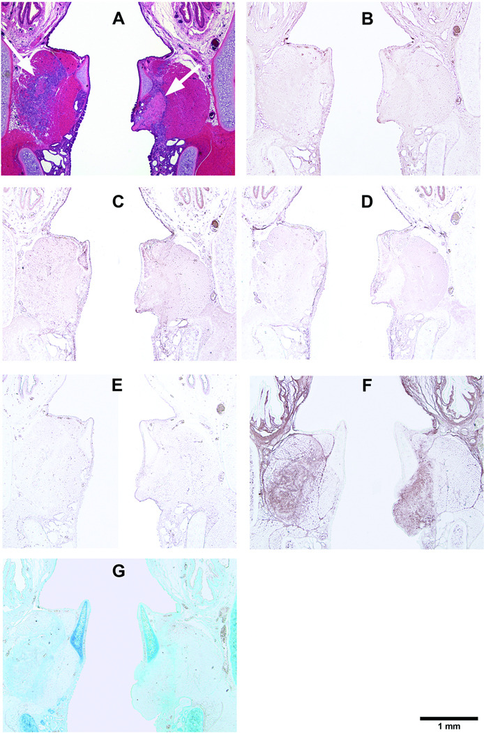
Figure 4.
Histological coronal sections of rat larynges showing HGF-loaded acellular scaffolds implanted into the right vocal fold wounds 3 days after surgery, with control scaffolds without HGF implanted into the left vocal fold wounds. (A) H&E (arrows indicating the implants on both sides); (B) Collagen type I; (C) Collagen type III; (D) Elastin; (E) Fibronectin; (F) Hyaluronic acid; and (G) Glycosaminoglycans.

