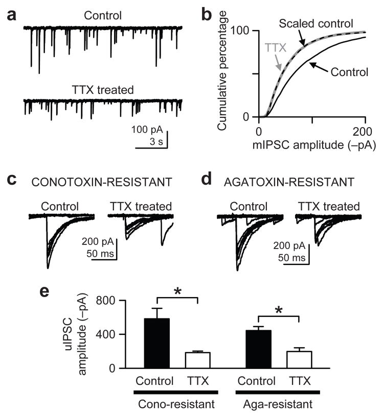
Figure 1.
Homeostatic downregulation of inhibitory synapses caused by chronic TTX treatment. (a) Whole-cell recordings of mIPSCs from CA1 pyramidal neurons in control and TTX-treated (3–5 days) slice cultures of rat hippocampus. Cells were voltage-clamped at −65 mV with a pipette solution containing 142.4 mM Cl−. (b) Cumulative distributions of mIPSCs (500 events per cell) show that chronic TTX reduced mIPSC amplitudes. The distribution of the scaled control data (dashed line), obtained after scaling down individual mIPSCs (see Methods), essentially overlapped exactly with the TTX-treated mIPSC distribution (thick gray line) (P > 0.9, Kolmogorov-Smirnov test between TTX and Scaled control), suggesting a global scaling of mIPSCs. (c) uIPSCs were evoked by extracellular minimal stimulation. Stimulus intensity was gradually increased until uIPSCs were elicited in an all-or-none fashion and consistently occurred, as shown in the sample traces. Slice cultures were preincubated with 500 nM ω-conotoxin GVIA for 15–30 min just before recording to isolate synapses that use P/Q-type Ca2+ channels for GABA release. (d) Same experiment as in c, but the slice cultures were preincubated with 300 nM ω-agatoxin IVA for 15–30 min. (e) Group data of uIPSC amplitudes show the mean amplitudes were reduced by chronic TTX in both conotoxin- and agatoxin-resistant synapses. *, P < 0.02. Error bars represent s.e.m. in this and all other figures.

