Figure 1.
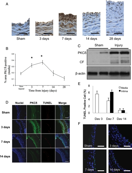
Expression of PKCδ and apoptosis in balloon-injured arteries. (A) Representative photomicrographs of balloon-injured rat carotid arteries stained for PKCδ (brown) 3, 7, 14, or 28 days post-injury. Sham-operated arteries serve as control (scale bar=50 µm). (B) Percentage of PKCδ positive area in the media by immunostaining (n = 5, *P < 0.05). (C) Balloon-injured rat carotid arteries were harvested 3 days after injury. Extracted proteins were subjected to immunoblotting for PKCδ. Beta-actin serves as a loading control. Each lane represents exatract from a single artery. (D) Representative photomicrographs of immunofluorescence staining for PKCδ (green), TUNEL staining (red), and nuclei (blue). Merged images are shown in the right panels (scale bar=100 µm). (E) TUNEL index were calculated as a ratio of the number of TUNEL-positive cells to total cell number (n = 6). (F) Representative photomicrographs of immunofluorescence staining for cleaved caspase-3 (green) (scale bar=20 um).

