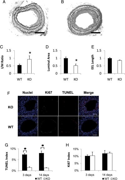
Figure 3.
Effects of PKCδ gene deficiency on intimal hyperplasia after carotid ligation. (A and B) Representative photomicrographs of the carotid arteries of PKCδ knock-out mice (KO) and their wild-type littermates (WT) 28 days after ligation. Sections were stained with Elastic-van Gieson staining (scale bar=50 µm). (C–E) Quantitative morphological analyses of the carotid artery of WT and KO 28 days after injury. The intimal area to medial area ratio (I/M ratio) (C), luminal area (D), and length of external elastic lamina (EEL) (E) were measured as described in the Methods (n = 6, *P < 0.05 as compared with WT mouse). (F) Representative micrographs of immunofluorescence staining of the carotid arteries of PKCδ knock-out mouse (KO) and their wild-type littermates (WT) 14 days after ligation. The cross sections were stained for nuclei (blue), Ki67 (green), and TUNEL (red). Merged images are shown on the right panels (scale bar=50 µm). TUNEL index (G) and Ki67 index (H) were calculated as a percentage of the number of positive cells to the number of total cells (n = 6, *P < 0.05 as compared with WT).

