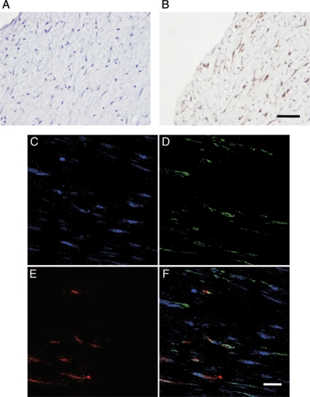
Figure 6.
PKCδ expression and apoptosis in human restenostic lesions. Representative micrographs of negative control (A) and immunostaining for PKCδ (B) of atherectomy specimens of human restenotic lesions (scale bar=50 µm). (C–E) Representative photomicrographs of double immunofluorescence staining for PKCδ (D: green), TUNEL staining (E: red), nuclei (C: blue), and merged image (F) (scale bar=20 µm).

