Figure 1.
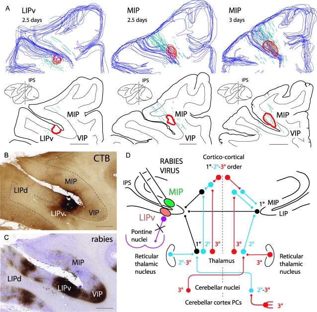
(A) 3D reconstructions and coronal sections of the injection area (red outlines) into the LIPv or the MIP at the level of the middle rostrocaudal third of the IPS, visualized by CTB immunolabeling at 2.5 or 3 days after injection of a mixture of rabies and CTB. Blue outlines: cortical surface. Green outlines: reconstructions of electrode tracts from previous recordings. (B, C) Photomicrographs of adjoining sections at the LIPv injection site, immunolabeled for CTB and rabies virus (same level as in A, left). Note that the injection area is easily identifiable with CTB (B) but not with rabies virus (C) at 2.5 days because of strong rabies immunolabeling of short-distance projections neurons in the IPS. In (B), shading in the white matter ventral to LIPv is background staining of an area of fibrosis due to multiple recording traces in that region. (D) Summary of the pathways of rabies retrograde transneuronal transfer to the cerebellar nuclei after coinjection of rabies virus and CTB into ventral MIP or LIPv: 1° (black), first-order neurons (conventional tracer, CTB) in the ipsilateral thalamus and cortical areas (ipsilateral corticocortical inputs, callosal inputs from homotopic IPS areas); 2° (blue), second-order neurons labeled transneuronally (rabies virus) at 2.5 days in the contralateral cerebellar nuclei, in the ipsilateral thalamic nuclei and reticular thalamic nucleus, and in the contralateral thalamic nuclei (the latter reflecting projections to IPS areas of the right hemisphere); 3° (red), third-order neurons labeled at 3 days in the contralateral cerebellar cortex (PCs) and contralateral reticular thalamic nucleus. Anterograde transneuronal transfer (e.g., to the pontine nuclei) did not occur (violet). Scale bar = 4000 μm in (A), 2000 μm in (B) (also applies to C).

