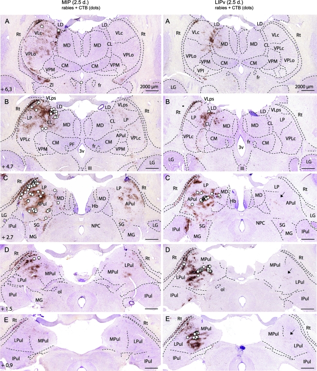
Figure 2.
Differences in thalamic (direct and second-order) inputs to the MIP (left) versus the LIPv (right) at 2.5 days after coinjection of rabies virus and the conventional tracer CTB. In each row, the left side is ipsilateral. The rostrocaudal distance of the coronal sections from the interaural axis is indicated. White dots: first-order neurons (CTB); note the topographical differences of neurons targeting MIP (dorsal and lateral) versus LIPv (caudal, medial, and ventral). Location: pulvinar complex, LP, VLps and VLc, CL, MD. Brown: rabies retrograde transneuronal labeling at 2.5 days, involving second-order neurons in the reticular thalamic (Rt) nucleus ipsilaterally and additional thalamic nuclei on both sides. Second-order neurons (brown) contralaterally (for LIPv, see arrows in C–E) mirror the distribution of first-order neurons (white dots) ipsilaterally. In both MIP and LIPv experiments, no labeling is found in Rt (third order) contralaterally, showing that third-order neurons are not yet labeled at 2.5 days (see also Fig. 1D). Other abbreviations: CM, centromedian; fr, fasciculus retroflexus; Hb, habenula; IPul, inferior pulvinar; LD, lateral dorsal; LG, lateral geniculate; MG, medial geniculate; MPul, medial pulvinar; NPC, nucleus of the posterior commissure; ol, olivary pretectal nucleus; PF, parafascicular; SG, suprageniculate; VPLc, ventral posterior lateral, pars caudalis; VPLo, ventral posterior lateral, pars oralis; VPM, ventral posterior medial; ZI, zona incerta; 3v, third ventricle; III, oculomotor nucleus.

