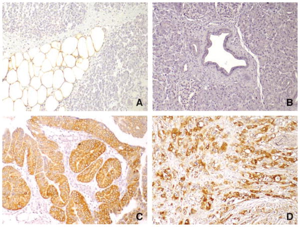
Figure 3.
Immunohistochemical analysis of FAS protein expression in tissue microarrays. A. Normal pancreatic adipose tissue with strong labeling of FAS. B. Normal pancreatic ductal cells and surrounding stromal cells do not label for FAS. C. IPMN is strongly positive for FAS in the neoplastic pancreatic ductal cells. D. Pancreatic ductal adenocarcinoma cells are strongly positive for FAS.

