Figure 5.

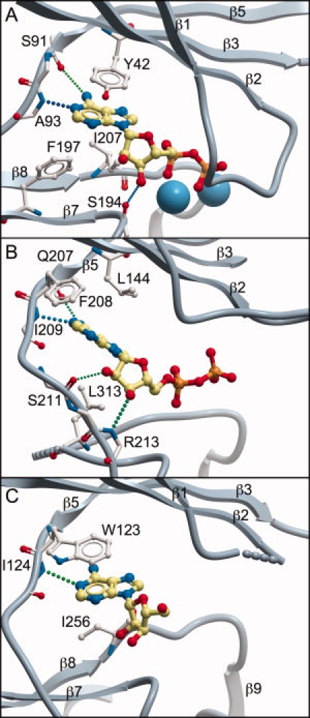
Comparison of the adenosine binding pocket in YtaA and other CAKs. Structures are presented in identical orientation, based on structural alignment and superposition with DaliLite.30 This presentation highlights changes in location and orientation of the adenosine molecule in each structure. Key adenosine-interacting residues in each structure are shown (side chains are omitted for residues that contribute only backbone atoms). H-bonds are shown as dotted lines and metal atoms as blue spheres. Portions of the structures are omitted to improve clarity. A: APH; B: ChoK; and C: YtaA.
