
Figure 7.
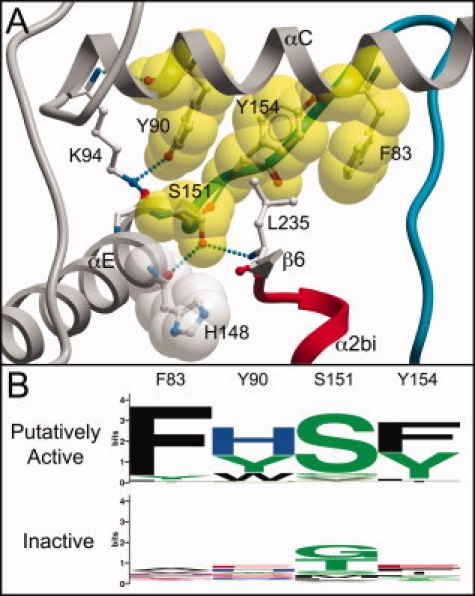
The hydrophobic linker motif conserved only in putatively active BSKs. A: Cutaway view of motif residue interactions. The YtaA structure is in approximately the same orientation as in Figure 4, with secondary structure elements identically colored. Motif residues are rendered with a yellow space-filling shell. H148, a residue highly conserved in almost all PKL kinases,11 is shown with a space-filling shell in white. Unconserved residues interacting with the motif are in ball-and-stick view. B: Logo of motif, showing selective conservation only in putatively active BSKs. The motif is discontinuous in sequence, and shown with intervening sequence removed.