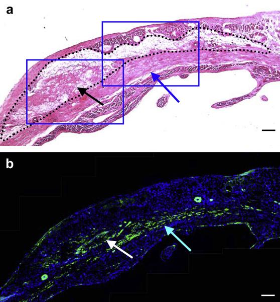
Figure 10.
Higher magnification of (a) H&E staining and (b) immunohistochemical staining in the hydrogel injected ventricular wall of sequential sections. Black dots indicate the injected hydrogel area and blue box indicates higher magnification area shown in Figure 11 (a). α-SMA staining appears green and nuclear staining appears blue (b). Black and white arrows in (a) and (b) respectively indicate tissue ingrowth in the hydrogel area, while blue arrows in (a) and (b) indicate smooth muscle populated area beneath the hydrogel area. Scale bars: 100 μm.

