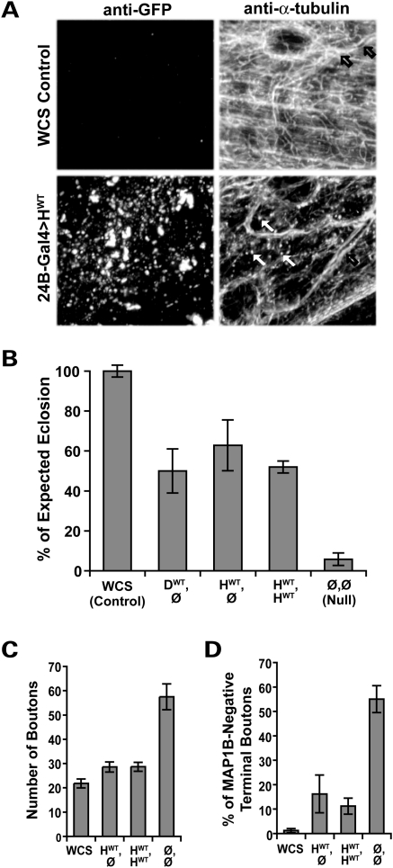
Figure 2.
Human and Drosophila Spastin are functionally equivalent. (A) Microtubules in the body wall muscles of wild-type third-instar Drosophila larvae were visualized using an antibody against α-tubulin, revealing a dense filamentous mesh throughout the tissue (top right). Microtubules in the walls of the trachea overlying the muscle are also seen (black arrows). Muscle-specific overexpression of human wild-type (HWT) Spastin revealed by anti-GFP (bottom left) causes an overall reduction in the intensity of α-tubulin staining (bottom right), as well as a profusion of punctae (white arrows). The punctate microtubule signal is distinct from the pattern of GFP expression, and likely reflects fragments resulting from the severing activity of the overexpressed human Spastin. (B) Pan-neuronal expression of fly or human spastin in the fly spastin null background rescues eclosion rates equivalently. Flies deleted for spastin (Ø,Ø) eclose successfully only ∼6% of the time, compared with WCS control flies, which nearly always eclose. GS-elav-GAL4-driven expression of a single copy of the fly or human spastin transgene (genotypes DWT,Ø and HWT,Ø, respectively) restores eclosion to over 50%, as does two copies of the human transgene (HWT,HWT; P < 5 × 10⁻⁶ for all groups compared with nulls, P > 0.4 between all transgenic lines). Similar results were observed for multiple independent insertions of the HWT transgene (Supplementary Material, Table S1). (C) At the cellular level, increased synaptic bouton number caused by loss of spastin is almost fully rescued by neuronal expression of the wild-type human transgene. Bouton number is 2.5-fold greater in spastin nulls (Ø,Ø) compared with WCS controls, and doubled compared with animals expressing human spastin (see also Fig. 3E). (D) Correspondingly, expression of human spastin significantly restores the penetration of microtubules into terminal synaptic boutons, such that only 16% (HWT,Ø) and 11% (HWT,HWT) are devoid of detectable MAP1B staining compared with >50% in nulls (P < 2 × 10⁻⁴ for either rescued genotype). WCS animals have <2% of terminal boutons lacking microtubules. Averages in this and subsequent figures are mean ± s.e.; P-values were calculated by one-way ANOVA; N's are detailed in the Methods.

