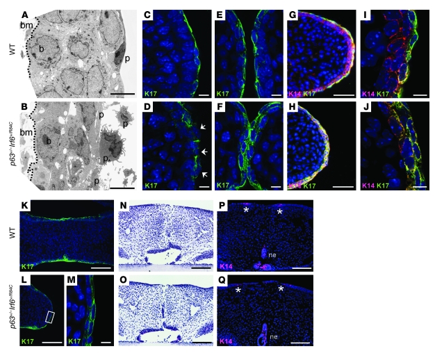
Figure 2. Cleft palate observed in p63+/–Irf6+/R84C mice.
(A and B) TEM analysis of the MEE at E14.5 revealed highly disordered basal and periderm cells in p63+/–Irf6+/R84C mice compared with their wild-type littermates. (C–J) Deconvolution analysis of the palatal shelves. (C and D) At E13.5, K17 appeared filamentous and incorrectly localized in the MEE (D, arrows). (E and F) At E14.5, whereas K17 expression was confined to the periderm of wild-type mice, K17 was expressed throughout the MEE in p63+/–Irf6+/R84C embryos. (G–J) K14 and K17 dual staining. (G and I) In wild-type mice, K14-positive basal cells were covered by K17/K14-positive periderm cells. (H and J) In p63+/–Irf6+/R84C embryos, the entire MEE stained positively for both K14 and K17. (K–M) At E15.5, whereas the MEE of wild-type mice degenerated, K17-positive cells persisted over the MEE of p63+/–Irf6+/R84C embryos. (M) Higher-magnification view of the boxed region in L. (N–Q) In vitro culture indicated that after 72 hours of forced contact, the palatal shelves of p63+/–Irf6+/R84C mice fused. K14 expression in both wild-type and p63+/–Irf6+/R84C palates was evident in remnants of the nasal and oral epithelia only (P and Q, asterisks). bm, basement membrane; p, periderm cell; b, basal cell; ne, nasal epithelium. Scale bars: 5 μm (A and B); 10 μm (C–F, I, J, and M); 50 μm (G and H); 100 μm (K, L, and N–Q).

