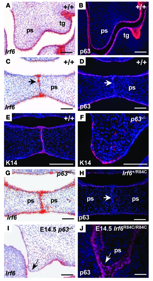
Figure 3. Irf6 and p63 expression during palatal development.
(A and B) At E13.5, Irf6 and p63 were expressed in similar domains in the epithelia of the oral cavity and in the tooth germs (tg) of wild-type mice. (C and D) At E14.5, Irf6 transcripts were strongly expressed in the midline seam and epithelial triangles (C, arrow); in contrast, p63 protein levels were downregulated in the MEE (D, arrow). (E and F) Immunofluorescence assays using K14 indicated that the palatal shelves of p63–/– mice, which exhibited a thin and fragile epithelium, were competent to express this protein. (G and H) Analysis of p63 and Irf6 in Irf6+/R84C and p63+/– embryos, respectively. Expression was unchanged in the heterozygous animals. (I) In E14.5 p63–/– palatal shelves, Irf6 transcripts were downregulated in the MEE (arrow). (J) p63 expression was maintained throughout the MEE of E14.5 Irf6R84C/R84C palatal shelves (arrow). Scale bars: 100 μm.

