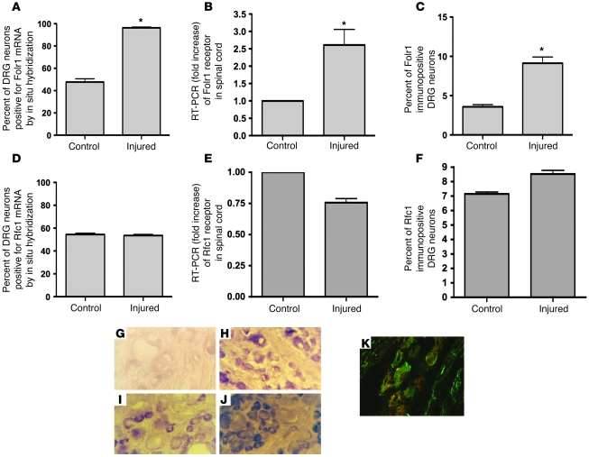
Figure 2. Combined spinal cord and peripheral nerve injury induces expression of Folr1 but not Rfc1.
4 days after sharp transection of both dorsal columns and the left sciatic nerve in rats, the left L5 and L6 DRGs were removed, sectioned, and mounted to undergo in situ hybridization (A, D) and immunohistochemistry (C, F) of the Folr1 and Rfc1 receptors. The spinal cord was removed for RT-PCR (B, E) and Western immunoblot (Figure 7, A and E). Note the significant upregulation of Folr1 with injury, with no change in the Rfc1 levels both in the spinal cord (SC) and DRG: in situ Folr1 n = 8 (SC), 8 (DRG); in situ Rfc1 n = 5 (SC), 5 (DRG); Folr1 RT-PCR n = 3 (SC), 3 (DRG); Rfc1 RT-PCR n = 3 (SC), 3 (DRG); immuno Folr1 n = 4 (SC), 4 (DRG); immuno Rfc1 n = 4 (SC), 4 (DRG). 2-tailed Student’s t test; mean ± SEM; *P < 0.05. (G, H) Folr1 in situ hybridization of DRG sections without (G) and with (H) preceding combined spinal cord and left sciatic nerve injury. (I, J) Folr1 immunostaining of DRG sections without (I) and with (J) preceding combined spinal cord and left sciatic nerve injury. (K) Confocal microscopy showing colocalization of the red Folr1 stain with the green neuronal (neurofilament) stain. Original magnification, ×40.

