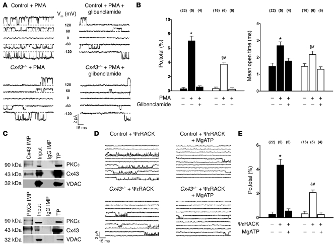
Figure 5. PKC-mediated stimulation of mitoKATP channel activity is diminished in Cx43+/– mice.
(A) MitoKATP single-channel activity is activated by PKC stimulation with PMA 2 μM (top left), which can be inhibited by glibenclamide 10 μM (top right). PMA-induced mitoKATP channel activation is reduced in Cx43+/– mice (bottom). Voltages are indicated. (B) Mean values of mitoKATP channel open probability (Po,total; left) and mean open time (right) in control (black) and Cx43+/– mice (white) in the absence and presence of PMA and glibenclamide, as indicated; n values are shown in parentheses. *P < 0.05 versus control; §P < 0.05 versus Cx43+/–; #P < 0.05 versus control + PMA. (C) Proteins of mitochondria were immunoprecipitated (IMP) for Cx43 (top) or PKCε (bottom). Western blot analysis for coprecipitated Cx43 and PKCε revealed positive results. Precipitation of mitochondrial proteins with anti-rabbit IgGs was used as negative control. Input lysates and a total ventricular protein extract (TP) were analyzed as positive control. Immunoblotting against VDAC was performed to exclude any unspecific coimmunoprecipitation. (D) MitoKATP single-channel activity was activated by selective PKCε stimulation with ψεRACK (top left), which could be inhibited by MgATP (top right). ψεRACK-induced mitoKATP channel activation was reduced in Cx43+/– mice (bottom). (E) Mean values of mitoKATP channel open probability in control (Po,total; black) and Cx43+/– mice (white) in the absence and presence of the selective PKCε peptide agonist ψεRACK (0.5 μM) and MgATP (100 μM), as indicated; n values are shown in parentheses. *P < 0.05 versus control; §P < 0.05 versus Cx43+/–; #P < 0.05 versus control + ψεRACK.

