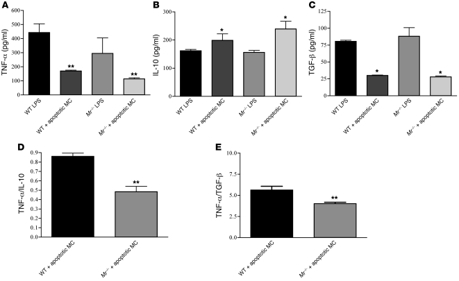
Figure 6. Cytokine release by WT and Mr–/– bone marrow–derived macrophages after stimulation with LPS and apoptotic WT MCs.
Bone marrow–derived macrophages were incubated with 0.5 μg/ml LPS with or without apoptotic WT MCs. (A–C) Cultured supernatants were harvested 2 hours after macrophage stimulation and assessed for TNF-α (A), IL-10 (B), and TGF-β (C). WT and Mr–/– macrophages reduced TNF-α production after uptake of apoptotic MCs, but the effect was significantly greater in Mr–/– cells. (D and E) TNF-α/IL-10 (D) and TNF-α/TGF-β (E) ratios confirmed a more significant noninflammatory phenotype in Mr–/– than in WT cells. *P < 0.05; **P < 0.01.

