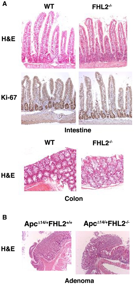
Figure 1. Histological analysis of normal intestine in FHL2−/− mice and intestinal adenomas in ApcΔ14/+FHL2−/− mice.
A. Normal intestinal architecture in FHL2−/− mice. Intestine and colon sections from 11 month-old wt and FHL2−/− mice were stained with H&E or Ki-67 by immunostaining. B. Intestinal adenomas in ApcΔ14/+FHL2+/+ and ApcΔ14/+FHL2−/− mice. Original magnifications, X100 (A and B).

