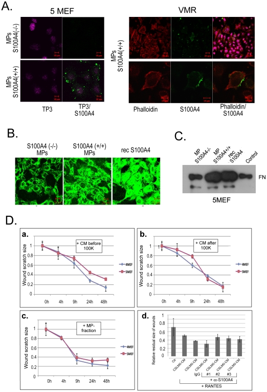
Figure 4. Functional significance of extracellular S100A4.
(A) Distribution of microparticles isolated from S100A4-positive 4MEF cells added to 5MEF and VMR cells. Immunofluorescence staining was performed with anti-S100A4 antibodies (green), rhodamine phalloidin (red), and nuclear staining with TO-Pro (TP3) (pink). (B) Immunofluorescence detection of FN (green) in 5MEF cells in response to S100A4+/+ and S100A4−/− microparticles from 4MEF and 5MEF cells, and 1µg/ml of the recombinant oligomeric S100A4 protein, respectively. (C) Detection of FN by Western blot analysis of cell lysates from 5MEF treated with S100A4−/− and S100A4+/+ microparticles and recombinant S100A4. As a control cell lysate from non-treated cells were used. FN band corresponding to molecular weight of approximately 250 kDa is indicated. (D) Effects of S100A4 microparticles on wound healing in 5MEF cells. Conditioned media from 4MEF and 5MEF cells before (a) and after (b) 100,000×g centrifugation and isolated microparticles (c) were added to scratched monolayers of 5MEF cells. Time-course kinetics of residual wounds are depicted in the graphs. (d) Wound healing assay with 4MEF cells. The residual size of scratches 12 h after “healing” is presented. Three different batches (#1, 2 and 3) of affinity purified polyclonal anti-S100A4 antibodies were used.

